Frozen Tissues
Frozen tissue is a valuable resource for research and diagnostics. Snap freezing helps maintain cellular structure and the integrity of biomolecules, such as DNA, RNA, and proteins, allowing for accurate analysis of disease mechanisms at both histological and molecular levels. Cryostat sections of frozen tissue are used for histological quality control.
Ultra-Low Freezers (-80°C)
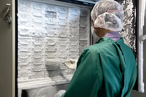
Ultra-low temperature freezers (-80°C) are effective for medium- to long-term storage (months to a few years) of human tissue samples, especially for preserving DNA, RNA, proteins, and other molecular components. They are widely used in research and clinical labs because they are cost-effective and easy to manage compared to liquid nitrogen storage.
However, they are not ideal for extremely long-term storage (decades) or for preserving live cell viability, as gradual degradation and ice crystal formation can occur over time. For extremely long-term storage or live cells, liquid nitrogen storage (-196°C) is preferred.
References
Shabihkhani, M., et al. (2014). The procurement, storage, and quality assurance of frozen blood and tissue biospecimens. Clinical Biochemistry, 47(4-5), 258–266.
Hubel, A., Spindler, R., & Skubitz, A. P. (2014). Storage of human biospecimens: selection of the optimal storage temperature. Biopreservation and Biobanking, 12(3), 165–175.
Jewell, S. D., et al. (2002). Analysis of the molecular quality of human tissues: an experience from the Cooperative Human Tissue Network. American Journal of Clinical Pathology, 118(5), 733–741.

Liquid Nitrogen Freezers (-196°C)
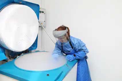
Liquid nitrogen freezers are among the best options for long-term storage of human tissue samples, particularly when it is essential to maintain cellular and molecular integrity for extended periods (decades or longer). These freezers operate at extremely low temperatures (-196°C), which makes them ideal for preserving both the structural integrity and biological function of tissues.
At -196°C, biochemical processes that lead to tissue degradation essentially stop, preserving DNA, RNA, proteins, and lipids almost indefinitely.
One distinction in liquid nitrogen storage is the difference between vapour phase and liquid phase storage. Vapor phase storage minimizes the risk of cross-contamination between samples, whereas liquid phase storage provides a more stable temperature but can increase contamination risks.
The low temperatures in liquid nitrogen prevent the formation of damaging ice crystals, which can disrupt cell membranes. This makes them suitable for storing tissues where cell viability is crucial, such as fertility preservation, regenerative medicine, and cryopreservation of living cells.

Flash Freezing
Also known as snap-freezing, this method involves rapidly freezing the tissue by immersing it in liquid nitrogen or placing it on dry ice. The temperature drop happens almost instantly.
Advantages
- Quick and Simple: Flash freezing is much faster and more convenient than controlled-rate freezing. It does not require specialized equipment beyond liquid nitrogen or dry ice.
- Good for Structural Preservation: Flash freezing is effective at preserving the molecular structure and chemical composition of tissues. It is especially useful for preserving tissues for biochemical assays (e.g., protein, DNA, RNA, or enzyme studies).
- Minimal Ice Crystal Formation in Small Samples: When done correctly, flash freezing can avoid the formation of large ice crystals, making it suitable for preserving small tissue samples.
- Cost-Effective: Requires less equipment and infrastructure, making it more affordable.
Disadvantages
- Not Ideal for Live Cells: Flash freezing often causes extensive ice crystal formation in large or delicate samples, damaging cell membranes and leading to poor cell viability. It’s not suitable for tissues that need to remain viable.
- Ice Crystals in Larger Samples: In larger tissues, rapid freezing can cause ice crystals to form within the tissue, potentially disrupting cellular structures.
Applications
- Biochemical and molecular assays (e.g., preserving tissues for RNA, DNA, or protein extraction).
- Tissues where cell viability is not required, but molecular integrity is crucial.
- Laboratories needing a quick and cost-effective method for freezing.
Controlled-Rate Freezing
This method involves gradually lowering the temperature of the tissue at a controlled, slow rate, typically using a specialized freezing machine (e.g., a controlled-rate freezer).
Advantages
- Prevents Ice Crystal Formation: By lowering the temperature gradually, controlled-rate freezing minimizes the formation of large ice crystals, which can damage cellular structures. This is particularly important for tissues containing delicate cells (e.g., live cells, embryos, or certain organs). The use of cryoprotectants, such as DMSO, further protects cells from damage by ice crystals.
- Optimal for Cell Viability: It is the preferred method when preserving live cells or tissues that may need to be revived later (e.g., stem cells or reproductive tissues).
- Precise Control: Controlled-rate freezing provides precise temperature management, which is beneficial when freezing samples that require specific freezing profiles.
Disadvantages
- Time-Consuming: Controlled-rate freezing is slower and requires specialized equipment, which may not be available in all laboratories.
- More Costly: The equipment and the time required make this method more expensive than flash freezing.
- Less Effective for Certain Molecules: For certain biochemical studies (like preserving proteins, RNA, or DNA), controlled-rate freezing may not be as necessary, as these molecules are often less sensitive to ice crystal formation.
Applications
- Preserving live tissues, cells, or organ samples where viability is important.
- Cryopreservation of embryos, oocytes, or stem cells.
- Samples where you want to avoid ice damage that could interfere with functionality.
Preparing Cryostat Sections
Cutting cryostat sections involves slicing frozen tissue samples into thin layers, typically using a cryostat, for use in various research and diagnostic applications. These sections are invaluable because they allow for the preservation of cellular and molecular integrity. The ability to maintain tissue morphology and biomolecular content in a near-native state makes cryostat sections essential for studying disease processes, identifying biomarkers, and validating therapeutic targets.
Best practices for cryostat sectioning include minimizing condensation buildup during handling to avoid tissue damage and using sharp blades to ensure clean sections. Proper labeling of samples is crucial for tracking tissue integrity over time.
The video opposite from Damien Harkin of the Queensland University of Technology (QUT), shows the preparation of frozen (or cryostat) sections.

